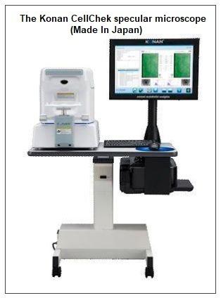
Standard biomicroscopy does not provide the critical detail of the earliest signs of endothelial disease. At Galbrecht EyeCare we use the CellChek Specular Microscope which allows us 100 x more magnification.
Specular microscopy is an important part of the testing process for all of our patients needing cataract surgery. Dr. Galbrecht explains, “This instrument is invaluable when determining if a patient is a good, great, or bad candidate for refractive surgery and cataract surgery. Most patients do not know that you must have a healthy endothelium to undergo any type of surgery to the eye.”
The CellChek Specular Microscope represents the latest in specular microscopy providing robust clinical evidence, and patented analysis methods that can reliably assess even problematic endothelium. It’s also fully automated with a wealth of features including auto-align, auto-focus, auto-capture, auto-analysis, and auto-pachymetry.
The CellChek captures high quality images of your corneal endothelium using a patented method that identifies the position of the cellular interface. It provides five focus points for image capture at the center and four peripheral sites, allowing a more comprehensive look at the cornea. This is particularly valuable in cases such as keratoconus, corneal transplantations, or corneal dystrophies.
Using non-contact optical pachymetry, the CellChek provides corneal thickness measurements at all five data sample sites. It’s an effective tool in showing patients the dangers of over wearing their contact lenses. “Many patients do not see the harm in sleeping in their contact lenses. This test shows the direct correlation for the amount of time spent sleeping in contact lenses and the damage it causes. The education given should be a warning to the patient about how to better care for their eyes”, explains Dr. Galbrecht.
The CellChek includes strong trends analysis to make robust use of minimum numbers of observable cells with advanced disease state corneas. It automatically records the location from which the data samples were acquired. This allows accurate re-assessment of specular data sample areas to trend cellular statistics over time, such as being able to assess and quantify over time, change in the cornea. Trends analysis is critical to to choosing treatment responses and understanding the progression or arrest of disease.
This test is also needed for patients contemplating implantation of phakic intraocular lenses or refractive surgery. Dr. Galbrect adds that “Without an adequate endothelial cell count, surgery is contraindicated. The same testing criterion for endothelial cell count is being implemented for refractive surgery as well. Since a successful LASIK procedure requires adequate thickness after the procedure is performed,”