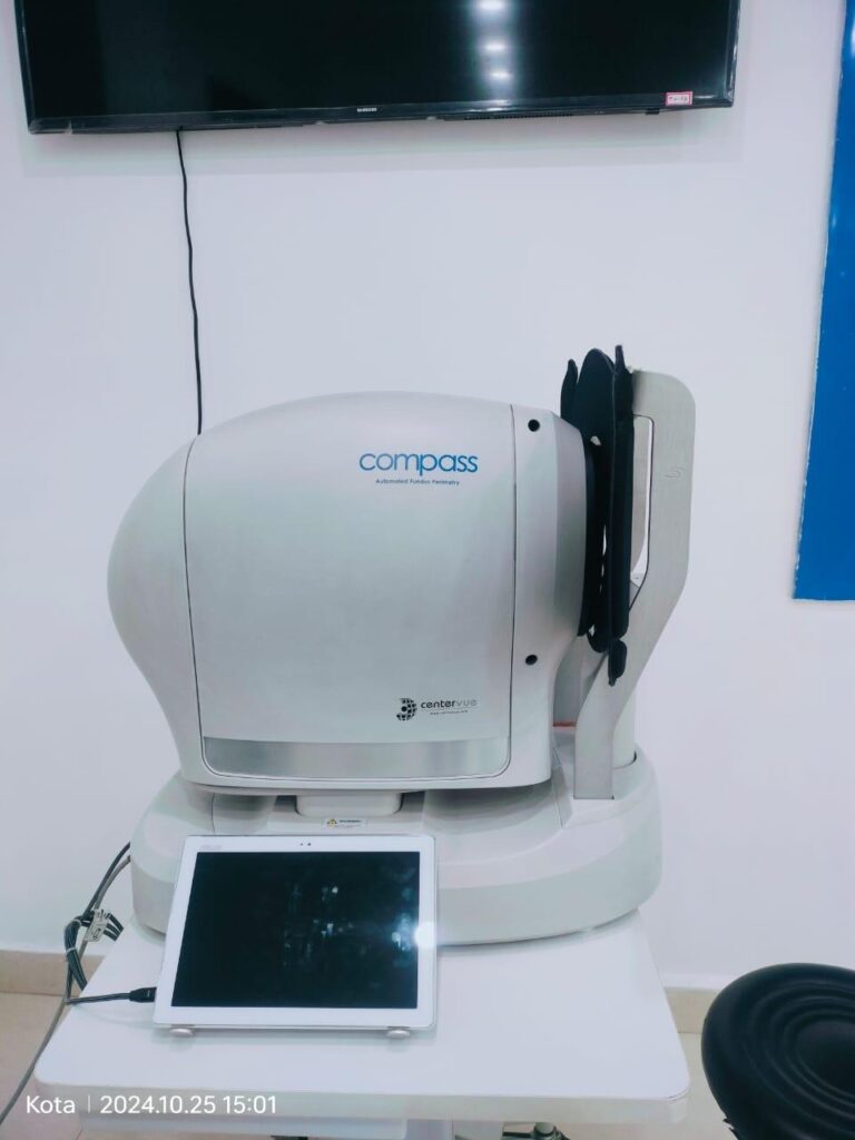
Fundus Automated Perimetry
COMPASS is the first Fundus Automated perimeter capable of performing standard 24-2 visual field testing, delivering TrueColor confocal images at the same time. COMPASS is a scanning ophthalmoscope combined with an automatic perimeter that provides confocal images of the retina, as well as measurements of retinal threshold sensitivity, under non-mydriatic conditions. Fundus automated perimetry is a technique that images the retina during visual field testing, enabling a correlation to be made between visual function and retinal structure.1 Advantages of Fundus Automated Perimetry over Standard Automated Perimetry include the possibility to measure sensitivity at specific retinal locations, higher accuracy thanks to retinal-tracking based compensation of eye movements and the simultaneous assessment of function (expressed by retinal sensitivity) and structure (images of the ONH, of the RNFL and of the retina). Fundus Automated Perimetry provides a simultaneous, quantitative assessment of fixation characteristics. Use of Fundus Automated Perimetry in the clinical management of glaucoma has been limited so far, as available systems were lacking compliance with the standards of automated perimetry. COMPASS overcomes such limitations and brings visual field analysis to the next level! In particular COMPASS, for the first time, extends field coverage to 30° + 30° and employs luminance parameters and a sensitivity scale as used in standard automated perimetry.
Benefits
Pupillary tracking
Key Features
High-resolution confocal imaging of the ONH and of the central retina
Compatibility to standard 24-2 visual field testing
As a perimeter, the system offers full compatibility with standard 24-2 visual field testing and contains an age-matched database of retinal sensitivity in normal subjects.
Superior quality of color and red-free images
As a retinal imager, COMPASS uses a confocal optical design, similarly to SLO systems, to capture color as well as red-free images of superior quality. In addition, a high resolution live image of the retina obtained using infrared illumination is available throughout the test.
Retinal tracking is at the heart of Fundus Automated Perimetry. Infrared images, acquired at the rate of 25 images per second, allow for continuous, automated, tracking of eye movements, with positional accuracy in the 10-20 microns range. Determination of eye movements yields to Fixation Analysis, where the location of the functional site of fixation and its stability are computed. Fixation analysis is unique to Fundus Automated Perimetry. Retinal tracking also yields to active compensation of fixation losses, with perimetric stimuli being automatically re-positioned prior to projection based on the current eye position. This mechanism is critical to reduce test-retest variability and ensure accurate correlation between function (i.e. retinal threshold values) and structure (retinal appearance). Compensation of eye movements takes place before and during the projection of a certain stimulus. In absence of this mechanism, a normal 2-3 degrees shift in eye position occurring at the time of projection of a certain stimulus would easily produce an artifact in VF results, with a wrong sensitivity being reported at that specific location.
Color confocal imaging SLO systems are superior to conventional fundus cameras in many ways, as they exploit a confocal imaging principle, which limits the effect of backscattered light from deeper layers and provides enhanced image quality in terms of contrast and resolution. Another major advantage of SLO systems is that they operate with much smaller pupils than non-confocal instruments. At the same time, though, SLO systems do not provide color images, as they typically employ monochromatic laser sources, resulting in black and white or pseudo-color images. Differently from existing SLO systems, Compass uses white light instead of monochromatic lasers, hence providing true color images and offering high fidelity to real retinal appearance. Compass images improve the diagnostic capabilities in the management of glaucoma as they offer: • no need for pupil dilation • excellent resolution and contrast • high quality even in presence of media opacities, such as cataract • optimized exposure of the ONH
At Sri Nirwana Netralaya, we believe that your vision is not just a sense, but a gift that deserves the highest level of care and attention. Our hospital is committed to preserving and enhancing this gift through a blend of cutting-edge technology, skilled professionals, and a compassionate approach to patient care.