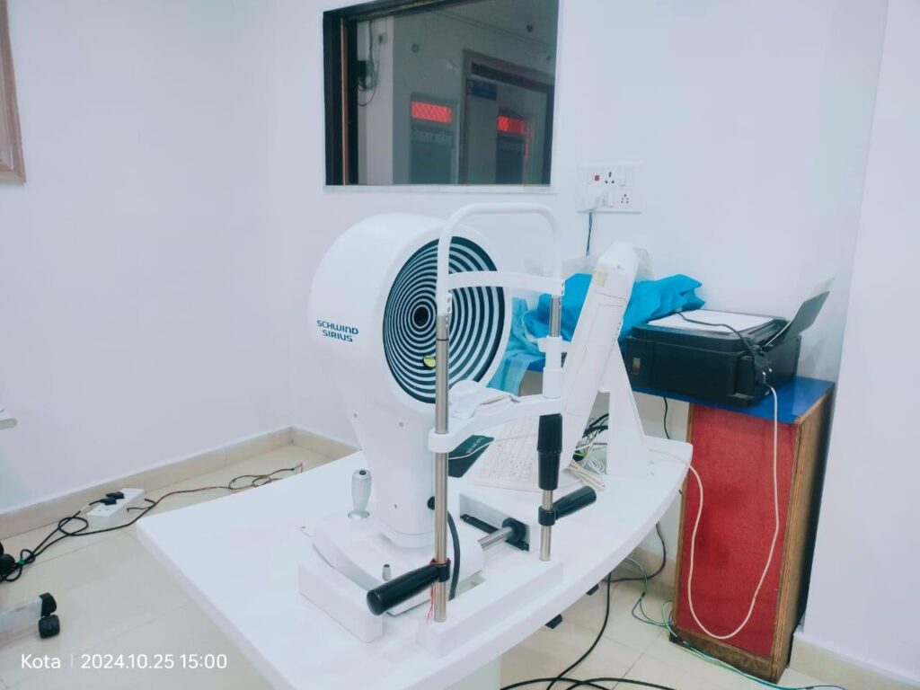
Precise data on the corneal thickness and corneal wavefront in one step
SCHWIND SIRIUS+ offers detailed information regarding corneal thickness and comprehensive support for corneal diagnosis.
The combination of a sophisticated 3D Scheimpflug camera and well-established Placido topography provides highly precise information about the entire anterior segment of the eye. All biometric measurements allow up to 100 corneal pachymetry sections for each acquisition.
Benefits at a glance
Precise information on pachymetry, elevation, curvature and dioptric power, each in relation to the anterior and posterior corneal surface
Diagnosis of the entire anterior segment of the eye in one step
High image quality over a corneal diameter of 16 millimetres
Calculation of all biometric measurements for the anterior segment of the eye with up to 100 high-resolution corneal pachymetry sections. The user can select the number of sections per measurement.
More precise centring and results through static cyclotorsion correction information for treatment with SCHWIND AMARIS and SCHWIND ATOS laser systems
Quick and precise
With 25 cross sections, SCHWIND SIRIUS+ records the entire anterior segment of the eye in about a second. The clever diagnostic system records detailed data on the corneal wavefront, topography of the anterior and posterior surface of the cornea, and the anterior chamber. The user can choose between particularly high image quality and particularly rapid measurement.
Perfect combined solution
SCHWIND SIRIUS+ uses the rotating 3D Scheimpflug camera to generate a pachymetry map of the eye and provide information about corneal thickness. The high-resolution Placido topography allows for a particularly detailed overview of the cornea including corneal aberrations. Here, the user can differentiate between the anterior, posterior and entire cornea. Maps and simulations help with analysis, as well as when talking to patients.
More benefits
Keratoconus screening
This convenient tool provides important corneal data and can make a pre-operative contribution to avoiding possible complications associated with ectasia.
IOL calculation
The calculation module for intraocular lenses, based on ray tracing technology, is excellently suited for treatment planning of refractively treated or untreated eyes.
Intrastromal rings
Placement of intrastromal rings can be planned exactly with the help of pachymetry maps and a corneal elevation profile.
Glaucoma screening
Various useful screening parameters assist glaucoma specialists with diagnosis
Pupillography
The integrated function captures the pupil diameter dynamically or statically depending on the defined lighting conditions. Precise data concerning the pupil centre and diameter are essential for planning and performing most refractive treatments.
Video keratoscopy
Special light sources for the stimulation of fluorescein help with the fitting of rigid contact lenses
“Dry eye” analyses
The high-resolution colour camera allows for a full evaluation, including advanced analysis of the condition of the tear film, the meibomian glands, hyperaemia of the conjunctiva and limbus, and the size of the tear meniscus. The result is a detailed report with a comprehensive assessment of the patient’s corneal condition and a substantial support for diagnosis of Dry Eye disease